Posted on

Prognostic factors for cardiovascular events in elderly pati | CIA

Angiotensinogen and Risk of Stroke Events in Patients with Type 2 Diab | DMSO

Background

The burden of community-acquired pneumonia (CAP) ranks the first among all infectious diseases, especially in the elderly people, with higher morbidity and mortality, more comorbidities and complications, higher need for admission to intensive care unit (ICU) and rate of clinical failure (CF), and more medical expenses.1–5 Cardiovascular diseases (ischemic heart disease, stroke) are the world’s biggest killers, responsible for 27% of the world’s total deaths in 2019, according to the World Health Organization (WHO).6 The incidence of CAP and cardiovascular diseases significantly increases with advanced age.5,7 Previous study indicated that older patients hospitalized with pneumonia had fourfold increased risk of subsequent occurrence of acute cardiovascular events (CVEs) in the first 30 days after pneumonia.8 A clinical rule that stratifies the risk of cardiac complications in patients hospitalized for CAP revealed that older patients are at major risk of CVEs after pneumonia.9 A global systematic review and meta‐analysis of observational studies demonstrated that the overall rates of cardiac complications after CAP were 13.9%, and the rate of heart failure was 9.2%, arrhythmias 7.2%, acute coronary syndromes 4.5%, and stroke 1.7%.10 Meanwhile, the occurrence of CVEs complicated the course of hospitalization with CAP. Compared to CAP patients without CVEs, CAP patients with CVEs had higher rate of mechanical ventilation, more need to ICU admission, prolonged length of stay (LOS), higher rate of short-term and long-term mortality, and higher 30-day re-hospitalization.10–12 Therefore, the mutual interaction between these two diseases should arouse more attention of respiratory physicians and cardiologists.

Current studies mainly focus on the overall population of patients with CAP, yet rare data are for the certain elderly population.13 Accordingly, we performed a multicenter, retrospective study to evaluate the burden of CVEs during hospitalization and to explore the independent prognostic factors for the occurrence of CVEs and 30-day mortality in elderly patients with CAP.

Methods

Study Setting, Design and Participants

This study is a multicenter, retrospective research on hospitalized elderly patients with CAP from the CAP-China network. Data of patients aged 65 years or older were abstracted from 13 centers in seven cities in three provinces between January 1, 2014 and December 31, 2014 (details are made available in the study by Han et al4). The study was approved by the Human Subject Protection Program Institutional Review Board at China-Japan Friendship Hospital. Additional approval was obtained from the local institutional review board of each participating hospital. Patient consent was waived owing to the retrospective and observational study design.

Inclusion/Exclusion Criteria and CAP Definition

Inclusion criteria included (1) age ≥ 65 years; (2) one of the top five discharge diagnoses defined as CAP. Exclusion criteria included (1) hospital-acquired pneumonia; (2) active tuberculosis; (3) non-infectious diseases, such as pulmonary infarction, tumor or pulmonary edema; (4) acquired immune deficiency syndrome; (5) re-admission within 72 hours after discharge.

CAP was defined as follows: (1) community onset; (2) presence of new infiltrate on chest X-ray or computed tomography scan together with at least one of the following: (i) new or increased cough (productive, non-productive or with a change in sputum characteristics) with or without dyspnea, chest pain or hemoptysis, (ii) fever, (iii) rales and/or signs of consolidation, (iv) peripheral WBC counts >10,000 cells·mm−3 or <4000 cells·mm−3, with or without a left shift toward immature forms.

Immunocompromised patients referred to these with solid-organ or stem cell transplant or bone marrow transplantation within one year of admission, chemotherapy for hematological disease or solid-tumor malignancy within six months of admission or neutropenia <500 cells·m−3, chest radiation therapy within one month of admission, prescription with immunosuppressive therapy within three months of admission or splenectomy.

Data Collection

Details on admission such as demographic data, a series of clinical information, hematological data, evaluation of initial antimicrobial treatment, are also available in the study by Han et al.4

Clinical failure (CF) is divided into early (≤72 hours) CF and late (>72 hours) CF. The detailed definitions are available in the study by Han et al.3

Definition of CVEs

The CVEs considered during the hospitalization were defined as follows:

  1. Events related with cardiac diseases: ① congestive heart failure (CHF) (new onset heart failure, or worsening preexisting heart failure with typical signs and/or symptoms associated with elevation of brain natriuretic peptide (BNP) or N-terminal pro-B-type natriuretic peptide (NT-proBNP);②new onset arrhythmia, or worsening preexisting arrhythmia (multifocal atrial tachycardia, atrial fibrillation or flutter; ventricular tachycardia, flutter or fibrillation; new onset of high degree atrio-ventricular block (2nd and 3rd));③ acute myocardial infarction (AMI) with typical signs or symptoms associated with troponin level above the normal value (according to the reference range values of local laboratory) and/or ischemic electrocardiographic changes (new ST-T changes or new left bundle branch block).
  2. Events related with cerebrovascular diseases (CVDs): new onset of hemorrhagic or ischemic stroke or transient ischemic attack defined as clinical manifestations and was confirmed by computed tomography or magnetic resonance imaging.
  3. Events related with thromboembolic diseases: pulmonary embolism (PE) or deep venous thrombosis (DVT) was confirmed by clinical manifestations and by pulmonary artery angiography computed tomography or eco-Doppler ultrasound, respectively.

All the patients diagnosed with CVEs (International Classification of Diseases, tenth revision [ICD-10] codes (in the Additional file 1: Table S1)) were based on clinical manifestations, laboratory tests and consultation with specialists.

Statistical Analysis

According to the occurrence of CVEs during hospitalization, the patients were divided into CVEs group and non-CVEs group. Categorical variables are presented as frequencies or percentages, and continuous variables are presented as median (interquartile range, IQR). The χ2 test is used for categorical variables and the Mann–Whitney U-test for continuous variables.

Variables showing significant difference in univariate analysis (p<0.10) are included in multivariate logistic regression analysis model for the occurrence of any CVEs and 30-day mortality in elderly patients with CAP, and a stepwise forward model is used to select independent risk factor. The 95% confidence intervals (CIs) and level of significance are reported.

All data are analyzed with SPSS (version 20, IBM Corp., New York, USA); p<0.05 is considered statistically significant.

Results

Study Population and Clinical Characteristics

Excluding immunosuppressed patients, 2941 patients aged ≥ 65 years were finally analyzed. 13.7% (n=402) of elderly patients with CAP occurred CVEs during hospitalization. Among them, 80.3% (n=323) were with acute CHF, 25.9% (n=104) with arrhythmia, 8.2% (n=33) with AMI, 6.2% (n=25) with CVDs, 4.0% (n=16) with thromboembolic diseases (six with PE and 10 with DVT). 73.1% (n=294) of patients had any one of CVEs, 22.6% (n=91) had two types of CVEs, 4.2% (n=17) had three types of CVEs.

Clinical characteristics of patients with or without CVEs are provided in Table 1. Patients with CVEs during hospitalization were older, higher risk of aspiration, long-term bedridden confinement and more comorbidities, especially cardiovascular disease and CVDs. Compared with non-CVEs patients, the pneumonia severity in patients with CVEs was more severe, history of CAP in past one year and use of glucocorticoids during hospitalization were more common.
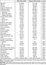
Table 1 Comparison of Clinical Characteristics in Hospitalized Elderly CAP Patients with or without CVEs (n=2941)

Clinical Manifestations, Laboratory and Radiologic Findings

Compared with non-CVEs patients, the rates of wheezing, cyanosis and lower extremity edema were more common in patients with CVEs, as well as unstable vital signs. Leukocytosis, hyperglycemia, azotemia, hyponatremia, hypoxemia, acidosis, hypoproteinemia, multilobe infiltration and pleural effusion were more prone to occur in patients with CVEs. During hospitalization, the incidence of related acute organ failure, diffuse intravascular coagulation (DIC) and gastrointestinal bleeding was notably different between the two groups (Table 2).
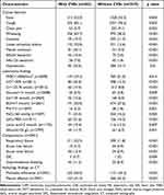
Table 2 Comparison of Clinical Manifestations, Laboratory and Radiologic Findings in Hospitalized Elderly CAP Patients with or without CVEs (n=2941)

Outcomes

Compared with non-CVEs patients, patients with CVEs during hospitalization were more prone to be admitted to ICU, administrated more guideline-discordant antibiotic therapy, higher rate of CF, and lower rate of clinical stability before discharge. The median LOS in patients with CVEs was 12 days, significantly longer than that (11 days) in non-CVEs patients (p=0.019). The in-hospital mortality and 30-day mortality were also significantly higher in CVEs patients than those in non-CVEs patients (p<0.001), 27.1% versus 2.0%, 30.3% versus 3.4% respectively. Median total cost for one elderly CAP patient with CVEs was RMB 20,315.5, significantly higher than that (RMB 12,207.1) for non-CVEs patient (p<0.001). All the data are provided in Table 1.

The occurrence of CVEs significantly increased with age (p<0.001), and the incidence in patients aged ≥ 86 years was as high as 25.9%, nearly fourfold than that in patients aged 65–70 years (Figure 1). The rate of CF, in-hospital mortality and 30-day mortality significantly increased with the numbers of CVEs (p<0.001). The rate of in-hospital mortality and 30-day mortality in patients with three types of CVEs was 64.7% and 70.6%, respectively, threefold than that in patients with one type of CVEs and 20–30 folds than that in patients without CVEs (p<0.001) (Figure 2).

Figure 1 The occurrence of CVEs, CF, in-hospital mortality, and 30-day mortality in elderly patients of different age groups. 1) As for the occurrence of CVEs, there are statistical differences between the two groups (p<0.05), except for the group aged 65–70 years and 71–75 years, group aged 76–80 years and 81–85 years. 2) As for the rate of CF, there are statistical differences between the group aged ≥86 years and all other groups (p<0.05). 3) As for the rate of in-hospital, there are statistical differences between the group aged 65–70 years versus group aged 81–85 years and group aged ≥86 years (p<0.05); There are statistical differences between the group aged ≥86 years and all other groups (p<0.05). 4) As for the rate of 30-day mortality, there are statistical differences between the group aged ≥86 years and all other groups (p<0.05).

Abbreviations: CVE, cardiovascular event; CF, clinical failure.

Figure 2 The occurrence of CF, in-hospital mortality, and 30-day mortality in elderly patients with different numbers of cardiovascular events. As for the rate of CF, in-hospital and 30-day mortality, there are statistical differences between the two groups (p<0.05), except for group with two and three types of CVEs.

Abbreviation: CF, clinical failure.

Predictive Factors for the Occurrence of CVEs During Hospitalization

Table 3 shows that previous history of CHF (OR 6.16; 95% CI, 4.14–9.18, p<0.001), CF (OR 4.69; 95% CI, 3.392–6.48, p<0.001), previous history of ischemic heart disease (OR 2.22; 95% CI, 1.61–3.07, p<0.001), use of glucocorticoids during hospitalization (OR 2.0; 95% CI, 1.39–2.89, p<0.001), aspiration (OR 1.88; 95% CI, 1.26–2.81, p=0.002), pleural effusion (OR 1.66; 95% CI, 1.25–2.20, p<0.001), multilobe infiltration (OR 1.50; 95% CI, 1.15–1.96, p=0.003), age (OR 1.05; 95% CI, 1.04–1.07, p<0.001), and blood urea nitrogen (BUN) (OR 1.03; 95% CI, 1.01–1.06, p=0.007) were independent predictors for the occurrence of any CVE during hospitalization in the multivariable logistic regression model. While level of blood sodium (OR 0.98; 95% CI, 0.97–0.99, p=0.007) was a protective factor.
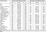
Table 3 Predictive Factors for the Occurrence of CVEs During Hospitalization in Univariate and Multivariable Logistic Regression Analysis

Predictive Factors for 30-Day Mortality in Elderly Patients with CAP

Table 4 shows that renal failure (OR 9.46; 95% CI, 4.17–21.48, p<0.001), respiratory failure (OR 9.32; 95% CI, 5.91–14.71, p<0.001), sepsis/sepsis shock (OR 7.87; 95% CI, 3.58–17.31, p<0.001), new CVDs (OR 5.94; 95% CI, 1.78–19.87, p=0.004), new heart failure (OR 4.04; 95% CI, 1.15–14.14, p=0.029), new arrhythmia (OR 2.38; 95% CI, 1.11–5.14, p=0.027), aspiration (OR 1.95; 95% CI, 1.09–3.50, p=0.025), CURB-65 (OR 1.57; 95% CI, 1.21–2.02, p=0.001), and white blood cell (WBC) count (OR 1.05; 95% CI, 1.02–1.09, p=0.006) were independent predictors for 30-day mortality in elderly patients with CAP in multivariable logistic regression model. While lymphocyte count (OR 0.63; 95% CI, 0.46–0.87, p=0.006) was a protective factor.
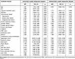
Table 4 Predictive Factors for 30-Day Mortality in Elderly CAP Patients in Univariate and Multivariable Logistic Regression Analysis

Discussion

This is the first retrospective multicenter study to evaluate the disease burden of CVEs and risk factors for incidence of CVEs in hospitalized elderly patients with CAP in China. Our study discovers that: 1) 13.7% of elderly patients with CAP experience CVEs during hospitalization with higher rate of CF and poorer prognosis. 2) Independent risk factors associated with CVEs are age, aspiration, previous history of CHF and ischemic heart disease, level of BUN, CF, use of systematic glucocorticoids during hospitalization, pleural effusion, and multilobe infiltration; while level of blood sodium is a protective factor. 3) Independent risk factors associated with 30-day mortality are renal failure, respiratory failure, sepsis/sepsis shock, new CVDs, new heart failure, new arrhythmia, aspiration, CURB-65, and WBC count; while lymphocyte count is a protective factor.

Nowadays, there were some researches on CVEs during the course of or after CAP. However, the incidence of CVEs differs as the different inclusion criteria of CVEs. In a multicenter prospective study enrolled 1266 patients with CAP, data indicated that 23.8% of patients experienced at least a CV event (excluded the thromboembolic diseases),12 higher than that in our study. We considered the gap is mainly related to the low proportion of patients in PSI class IV or V or CURB-65 class 3–5. The most common CVE during hospitalization is newly diagnosed acute heart failure (11.0%), followed by arrhythmia (3.5%) and AMI (1.1%), similar with the data from systematic review and meta-analysis of observational studies.10 As to the mechanisms about the occurrence of CVEs in patients with CAP, previous studies demonstrated acute or persistent inflammation after CAP became the fuse of potential triggers for cardiac events, leading to the increased pro-inflammatory cytokines or activation of pro-coagulant factors. Plaque-related CVEs (AMI) and plaque-unrelated CVEs (arrhythmias and heart failure) occurred after plaque rupture, in-situ thrombus formation, and alteration of the balance of arterial perfusion or diffuse organ abnormalities with cellular dysfunction.14–18 Additionally, the occurrence of type 2 myocardial infarction (T2MI) after acute infection is considered to be related to“demand ischemia” (a mismatch in myocardial oxygen supply and demand).19 In older adults, coronary stenosis from chronic plaques and possibly toxin-mediated vasoconstriction after acute infection may lead to increased cardiac metabolic mismatch.20 Data discovered T2MI gradually increased with advanced age, especially among patients aged over 75 years old.21 Thence, appropriate antimicrobial regimens, effective treatment against other underlying diseases, anticoagulation therapy, dynamic monitoring of laboratory indicators, and timely adjustment of therapeutic plans can play an important role in the control of inflammation.

The disease burden of elderly patients with CAP experienced CVEs is heavier. In a prospective multicenter cohort of 1182 CAP inpatients enrolled between 2011 and 2016, Francesco Violi et al reported patients who experienced a CVE were older, had a higher prevalence of underlying diseases and complications, higher disease severity, unstable vital signs and 30-day mortality,22 in line with our results. In our study, a significant increasing trend in the proportion of patients with CVEs, CF, and short-term mortality was found across the advanced age; meanwhile, the increased numbers of CVEs lead to poorer prognosis. Patients with CVEs had significantly lower proportion of guideline-concordant antibiotic therapy than that in patients without CVEs, thus, the proportion of CF significantly increased accordingly as well as prolonged LOS and higher short-term mortality.3 This result suggests that appropriated evaluation of pathogens plays an important role in the choice of optimal antimicrobial regimen.

We confirmed previous reports showing that history of heart failure, previously diagnosed coronary artery disease, pleural effusion, multilobe infiltration, age and BUN were independently associated with CVEs.12,16,23–26 In the first analysis of prospectively collected data from the Pneumonia Patient Outcomes Team cohort study in patients with CAP, sodium<130 mmol/L was considered as independent risk factor for incident cardiac complications.25 In our data, level of blood sodium (OR 0.98; 95% CI, 0.97–0.99, p=0.007) was protective factor for the occurrence of CVEs, suggesting the similar conclusion. Adjunctive use of corticosteroids for patients with CAP has been controversial, even for severe CAP.27–29 In a propensity-score adjusted Cox model by Cangemi et al, use of corticosteroid for patients with CAP was associated with a lower incidence of intra-hospital myocardial infarction (OR 0.46; 95% CI 0.24–0.88, p=0.02), albeit not reducing either overall mortality or cardiovascular death.30 Conversely, we found adjunctive use of corticosteroids during hospitalization was associated with a higher incidence of CVEs (OR 2.0; 95% CI, 1.39–2.89, p<0.001). In an observational study of 500 consecutive patients hospitalized with CAP who were enrolled in the Community-Acquired Pneumonia Organization (CAPO) cohort study, data addressed CF was significantly related to the occurrence of AMI (OR, 4.22; 95% CI, 1.10–16.29),31 generally consistent with our conclusion. 21.4% of elderly patients with CVEs in our population had higher risk of aspiration. Aspiration can bring about chemical pneumonia, bacterial pneumonia, or lipoid pneumonia, and thus lead to acute or chronic infection and inflammation. Acute infections not only destabilize vascular endothelium and result in an imbalance between myocardial oxygen supply and demand, but also have both systemic and local effects on coronary vessels, thence creating an increased risk of cardiovascular events.32,33 In our multivariable logistic regression analysis, aspiration was confirmed as an independent predictor for CVEs.

The results of the current study are concordant with previous evidence that aspiration, CURB-65, WBC count, sepsis/sepsis shock and respiratory failure were independent predictors for short-term mortality for elderly patients with CAP.4,34–37 We found the complication after CAP, renal failure was significantly associated with increased 30-day mortality (OR 9.46; 95% CI, 4.17–21.48, p<0.001). Yet, in the past literatures, BUN is more used to an evaluation indicator. A retrospective cohort study from China by Kang and coworkers assessing 4880 CAP patients aged ≥ 65 years showed that BUN was a prognostic factor for in-hospital mortality.35 Data from a multicenter prospective study on the Implications of acute Cardiovascular Events in patients hospitalized for Community-Acquired Pneumonia (ICECAP), enrolling patients consecutively hospitalized from 2016 to 2018, indicated the occurrence of any CVE during hospitalization independently and significantly increased the risk of 30-day mortality (HR 1.69; 95% CI, 1.14–2.51, p = 0.009); while newly diagnosed heart failure, new onset atrial fibrillation or flutter, acute coronary syndrome, separately, were not associated with increased risk of 30-day mortality.12 The current study is the first to our knowledge to illustrate new CVDs, new heart failure, and new arrhythmia were independently and significantly increased the risk of 30-day mortality for elderly patients with CAP.

There are some limitations in our study that should be acknowledged. The present study was a retrospective design; thus, missing data were inevitable There were no records about the timing of onset of CVEs, the detailed type of new arrhythmia and CVDs during hospitalization. Furthermore, medications like statins and anticoagulants during hospitalization were also not evaluated. Meanwhile, we did not evaluate the association between pathogens or antimicrobial treatment and the occurrence of CVEs. Finally, biomarkers such as D-dimer, BNP, NT-proBNP presented lots of missing values, thereby, the relationship between biomarkers and the occurrence of CVEs could not be explored.

Conclusions

CVEs during hospitalization are common in elderly patients with CAP in China. Patients with CVEs have heavier disease burden and poorer outcomes, especially those with new CVDs, new heart failure, and new arrhythmia, which are independently and significantly prognostic factors for short-term mortality. It is critical for clinicians to early identify risk factors and strengthen the hierarchical management of elderly patients with CAP.

Abbreviations

CVEs, cardiovascular events; CAP, community-acquired pneumonia; CHF, congestive heart failure; CVDs, cerebrovascular diseases; CF, clinical failure; OR, odds ratio; ICU, intensive care unit; WHO, World Health Organization; LOS, length of stay; BNP, brain natriuretic peptide; NT-proBNP, N-terminal pro-B-type natriuretic peptide; AMI, acute myocardial infarction; PE, pulmonary embolism; DVT, deep venous thrombosis; IQR, interquartile range; CIs, confidence intervals; DIC, diffuse intravascular coagulation; BUN, blood urea nitrogen; WBC, white blood cell; PSI, pneumonia severity index; T2MI, type 2 myocardial infarction; CAPO, Community-Acquired Pneumonia Organization; ICECAP, Implications of acute Cardiovascular Events in patients hospitalized for Community-Acquired Pneumonia; DIC, disseminated intravascular coagulation; COPD, chronic obstructive pulmonary disease; HCAP, healthcare-associated pneumonia; RR, respiratory rate; HR, heart rate; HCT, hematocrit; Cr, creatinine; Na, sodium; PaO2/FiO2, partial arterial oxygen pressure/fraction of inspired oxygen; PaO2, partial arterial oxygen pressure; SaO2, arterial oxygen saturation; CT, computed tomography.

Data Sharing Statement

All data generated or analyzed during this study are included in this published article and its supplementary information files.

Ethics Approval and Informed Consent

This study was approved by the China-Japan Friendship Hospital Ethics Committee (No. 2015–85) on October 12, 2015. We also confirmed that all patient data was treated with confidentiality, in accordance with the Declaration of Helsinki.

Consent for Publication

All authors have confirmed that the details of the paper.

Acknowledgments

The authors are grateful for the contributions of all the staff of the CAP-China network for their help with data collection and input. Thanks to Yimin Wang, Guangqiang Wang, Xuexin Yao, Hongxia Yu, Guohua Yu, Meng Liu, Chunxue Xue, Bo Liu, Xiaoli Zhu, Yanli Li, Ying Xiao, Xiaojing Cui, Lijuan Li, and Lei Wang for collecting the information. Thanks to Yi Wang for revising the figures.

Author Contributions

All authors made a significant contribution to the work reported, whether that is in the conception, study design, execution, acquisition of data, analysis and interpretation, or in all these areas; took part in drafting, revising or critically reviewing the article; gave final approval of the version to be published; have agreed on the journal to which the article has been submitted; and agree to be accountable for all aspects of the work.

Funding

This work was supported by the National Science Grant for Distinguished Young Scholars (grant number 81425001/H0104), the National Key Technology Support Program from Ministry of Science and Technology (grant number 2015BAI12B11) and the Beijing Science and Technology Project (grant number D151100002115004).

Disclosure

The authors declare that they have no competing interests.

References

1. Ferreira-Coimbra J, Sarda C, Rello J. Burden of community-acquired pneumonia and unmet clinical needs. Adv Ther. 2020;37(4):1302–1318. doi:10.1007/s12325-020-01248-7

2. Cillóniz C, Dominedò C, Pericàs JM, Rodriguez-Hurtado D, Torres A. Community-acquired pneumonia in critically ill very old patients: a growing problem. Eur Respir Rev. 2020;29(155):190126. doi:10.1183/16000617.0126-2019

3. Han X, Liu X, Chen L, et al. Disease burden and prognostic factors for clinical failure in elderly community acquired pneumonia patients. BMC Infect Dis. 2020;20(1):668. doi:10.1186/s12879-020-05362-3

4. Han X, Zhou F, Li H, et al. Effects of age, comorbidity and adherence to current antimicrobial guidelines on mortality in hospitalized elderly patients with community-acquired pneumonia. BMC Infect Dis. 2018;18(1):192. doi:10.1186/s12879-018-3098-5

5. Furman CD, Leinenbach A, Usher R, Elikkottil J, Arnold FW. Pneumonia in older adults. Curr Opin Infect Dis. 2021;34(2):135–141. doi:10.1097/QCO.0000000000000718

6. World Health Organization. The top 10 causes of death. Avialable from: https://www.who.int/news-room/fact-sheets/detail/the-top-10-causes-of-death. Accessed April 20, 2022.

7. Lopez-Otin C, Blasco MA, Partridge L, Serrano M, Kroemer G. The hallmarks of aging. Cell. 2013;153:1194–1217. doi:10.1016/j.cell.2013.05.039

8. Corrales-Medina VF, Alvarez KN, Weissfeld LA, et al. Association between hospitalization for pneumonia and subsequent risk of cardiovascular disease. JAMA. 2015;313:264–274. doi:10.1001/jama.2014.18229

9. Corrales-Medina VF, Taljaard M, Fine MJ, et al. Risk stratification for cardiac complications in patients hospitalized for community-acquired pneumonia. Mayo Clin Proc. 2014;89(1):60–68. doi:10.1016/j.mayocp.2013.09.015

10. Tralhão A, Póvoa P. Cardiovascular events after community‐acquired pneumonia: a global perspective with systematic review and meta‐analysis of observational studies. J Clin Med. 2020;9(2):414. doi:10.3390/jcm9020414

11. Chen L, Han X, Li Y, Zhang C, Xing X. Complications of cardiovascular events in patients hospitalized with influenza-related pneumonia. Infect Drug Resist. 2021;14:1363–1373. doi:10.2147/IDR.S305509

12. Pieralli F, Vannucchi V, Nozzoli C, et al. Acute cardiovascular events in patients with community acquired pneumonia: results from the observational prospective FADOI-ICECAP study. BMC Infect Dis. 2021;21(1):116. doi:10.1186/s12879-021-05781-w

13. Putot A, Bouhey E, Tetu J, et al. Troponin elevation in older patients with acute pneumonia: frequency and prognostic value. J Clin Med. 2020;9(11):3623. doi:10.3390/jcm9113623

14. Ross R, Epstein FH. Atherosclerosis – an inflammatory disease. N Engl J Med. 1999;340:115–126. doi:10.1056/NEJM199901143400207

15. Corrales-Medina VF, Madjid M, Musher DM. Role of acute infection in triggering acute coronary syndromes. Lancet Infect Dis. 2010;10:83–92. doi:10.1016/S1473-3099(09)70331-7

16. Aliberti S, Ramirez JA. Cardiac diseases complicating community-acquired pneumonia. Curr Opin Infect Dis. 2014;27(3):295–301. doi:10.1097/QCO.0000000000000055

17. Violi F, Carnevale R, Calvieri C, et al. Nox2 up-regulation is associated with an enhanced risk of atrial fibrillation in patients with pneumonia. Thorax. 2015;70:961–966. doi:10.1136/thoraxjnl-2015-207178

18. Rae N, Finch S, Chalmers JD. Cardiovascular disease as complication of community-acquired pneumonia. Curr Opin Pulm Med. 2016;22(3):212–218. doi:10.1097/MCP.0000000000000261

19. Musher DM, Abers MS, Corrales-Medina VF, Longo DL. Acute Infection and Myocardial Infarction. N Engl J Med. 2019;380(2):171–176. doi:10.1056/NEJMra1808137

20. Sibelius U, Grandel U, Buerke M, et al. Staphylococcal alpha-toxin provokes coronary vasoconstriction and loss in myocardial contractility in perfused rat hearts: role of thromboxane generation. Circulation. 2000;101(1):78–85. doi:10.1161/01.CIR.101.1.78

21. Putot A, Chague F, Manckoundia P, Cottin Y, Zeller M. Post-infectious myocardial infarction: new insights for improved screening. J Clin Med. 2019;8(6):827. doi:10.3390/jcm8060827

22. Violi F, Cangemi R, Falcone M, et al. Cardiovascular complications and short-term mortality risk in community-acquired pneumonia. Clin Infect Dis. 2017;64(11):1486–1493. doi:10.1093/cid/cix164

23. Aliberti S, Ramirez J, Cosentini R, et al. Acute myocardial infarction versus other cardiovascular events in community-acquired pneumonia. ERJ Open Res. 2015;1(1):00020–2015. doi:10.1183/23120541.00020-2015

24. Cangemi R, Calvieri C, Falcone M, et al. Relation of cardiac complications in the early phase of community-acquired pneumonia to long-term mortality and cardiovascular events. Am J Cardiol. 2015;116(4):647–651. doi:10.1016/j.amjcard.2015.05.028

25. Corrales-Medina VF, Musher DM, Wells GA, Chirinos JA, Chen L, Fine MJ. Cardiac complications in patients with community-acquired pneumonia: incidence, timing, risk factors, and association with short-term mortality. Circulation. 2012;125(6):773–781. doi:10.1161/CIRCULATIONAHA.111.040766

26. Viasus D, Garcia-Vidal C, Manresa F, Dorca J, Gudiol F, Carratalà J. Risk stratification and prognosis of acute cardiac events in hospitalized adults with community acquired pneumonia. J Infect. 2013;66(1):27–33. doi:10.1016/j.jinf.2012.09.003

27. Medical Letter on Drugs and Therapeutics. Corticosteroids in Community-Acquired Pneumonia. JAMA. 2020;323(9):887–888. doi:10.1001/jama.2020.0216

28. Jiang S, Liu T, Hu Y, et al. Efficacy and safety of glucocorticoids in the treatment of severe community-acquired pneumonia: a meta-analysis. Medicine. 2019;98(26):e16239. doi:10.1097/MD.0000000000016239

29. Torres A, Sibila O, Ferrer M, et al. Effect of corticosteroids on treatment failure among hospitalized patients with severe community-acquired pneumonia and high inflammatory response: a randomized clinical trial. JAMA. 2015;313(7):677–686. doi:10.1001/jama.2015.88

30. Cangemi R, Falcone M, Taliani G, et al. Corticosteroid use and incident myocardial infarction in adults hospitalized for community‐acquired pneumonia. Ann Am Thorac Soc. 2019;16(1):91–98. doi:10.1513/AnnalsATS.201806-419OC

31. Ramirez J, Aliberti S, Mirsaeidi M, et al. Acute myocardial infarction in hospitalized patients with community-acquired pneumonia. Clin Infect Dis. 2008;47(2):182–187. doi:10.1086/589246

32. Petroianni A, Ceccarelli D, Conti V, Terzano C. Aspiration pneumonia. Pathophysiological aspects, prevention and management. A review. Panminerva Med. 2006;48(4):231–239.

33. Singanayagam A, Singanayagam A, Elder DH, Chalmers JD. Is community-acquired pneumonia an independent risk factor for cardiovascular disease? Eur Respir J. 2012;39(1):187–196. doi:10.1183/09031936.00049111

34. Feng DY, Zou XL, Zhou YQ, Wu WB, Yang HL, Zhang TT. Combined neutrophil-to-lymphocyte ratio and CURB-65 score as an accurate predictor of mortality for community-acquired pneumonia in the elderly. Int J Gen Med. 2021;14:1133–1139. doi:10.2147/IJGM.S300776

35. Kang Y, Fang XY, Wang D, Wang XJ. Activity of daily living upon admission is an independent predictor of in-hospital mortality in older patients with community acquired pneumonia. BMC Infect Dis. 2021;21(1):314. doi:10.1186/s12879-021-06006-w

36. Yoshikawa H, Komiya K, Yamamoto T, et al. Quantitative assessment of erector spinae muscles and prognosis in elderly patients with pneumonia. Sci Rep. 2021;11(1):4319. doi:10.1038/s41598-021-83995-3

37. Leroy O, Bosquet C, Vandenbussche C, et al. Community-acquired pneumonia in the intensive care unit: epidemiological and prognosis data in older people. J Am Geriatr Soc. 1999;47(5):539–546. doi:10.1111/j.1532-5415.1999.tb02567.x

Posted on

Trajectories of prescription opioid dose and risk of opioid-related adverse events among older Medicare beneficiaries in the United States: A nested case–control study

Trajectories of prescription opioid dose and risk of opioid-related adverse events among older Medicare beneficiaries in the United States: A nested case–control study

Abstract

Methods and findings

We conducted a nested case–control study within a cohort of older (≥65 years) patients diagnosed with CNCP who were new users of prescription opioids, assembled using a 5% national random sample of Medicare beneficiaries from 2011 to 2018. From the cohort with a mean follow-up of 2.3 years, we identified 3,103 incident ORAE cases with ≥1 opioid prescription in 6 months preceding the event, and 3,103 controls matched on sex, age, and time since opioid initiation. Key exposure was trajectories of prescribed opioid morphine milligram equivalent (MME) daily dosage over 6 months before the incident ORAE or matched controls. Among the cases and controls, 2,192 (70.6%) were women, and the mean (SD) age was 77.1 (7.1) years. Four prescribed opioid trajectories before the incident ORAE diagnosis or matched date emerged: gradual dose discontinuation (from ≤3 to 0 daily MME, 1,456 [23.5%]), gradual dose increase (from 0 to >3 daily MME, 1,878 [30.3%]), consistent low dose (between 3 and 5 daily MME, 1,510 [24.3%]), and consistent moderate dose (>20 daily MME, 1,362 [22.0%]). Few older patients (<5%) were prescribed a mean daily dose of ≥90 daily MME during 6 months before diagnosis or matched date. Patients with gradual dose discontinuation versus those with a consistent low dose, moderate dose, and increase dose were more likely to be younger (65 to 74 years), Midwest US residents, and receiving no low-income subsidy. Compared to patients with gradual dose discontinuation, those with gradual dose increase (adjusted odds ratio [aOR] = 3.4; 95% confidence interval (CI) 2.8 to 4.0; P < 0.001), consistent low dose (aOR = 3.8; 95% CI 3.2 to 4.6; P < 0.001), and consistent moderate dose (aOR = 8.5; 95% CI 6.8 to 10.7; P < 0.001) had a higher risk of ORAE, after adjustment for covariates. Our main findings remained robust in the sensitivity analysis using a cohort study with inverse probability of treatment weighting analyses. Major limitations include the limited generalizability of the study findings and lack of information on illicit opioid use, which prevents understanding the clinical dose threshold level that increases the risk of ORAE in older adults.

Introduction

The number of older adults who had medical encounters for treatment of opioid misuse, dependence, and poisoning has increased disproportionately over the past decade [1]. The opioid-related adverse events (ORAEs) defined by the United States (US) government agencies [13] contain diagnostic codes commonly used for opioid use disorder (OUD) and overdose from use of illicit opioids (i.e., heroin) or incorrect use of prescribed opioids, as well as E codes for severe adverse effects from use of heroin or correct use of prescribed opioids that lead to hospital or emergency department visits. The rate of hospital stays and emergency department visits due to ORAEs rose by 34% (from 199.3 stays to 267.6 stays per 100,000 persons) and 74% (from 44.7 visits to 77.9 visits per 100,000 persons), respectively, among older patients between 2010 and 2015 [1]. A study of a commercially insured population also indicated a marked increase (14.2-fold from 2.05 to 31.12 per 10,000 persons) in the incidence of OUD or overdose among older adults aged 65 and older between 2006 and 2016 [4]. These alarming statistics have prompted questions of what might have predisposed older patients to be at risk for ORAEs [2].

Of the known risk factors, prescription opioid dose is one of the strong predictors of ORAEs [59]. Studies of nonelderly or mixed populations of young and older populations showed that use of prescription opioids at a dose of 90 morphine milligram equivalent (MME) or above per day was associated with an increased risk for opioid overdose and deaths [1013]. Built on this evidence, the 2016 Centers for Disease Control and Prevention (CDC)’s Guidance for Opioid Prescribing for Chronic Pain recommends avoidance of prescribing daily opioid doses at 90 mg MME or greater [14]. Since then, some medical societies’ guidelines [15], state regulations [16], and health insurance payers [17] have adapted the CDC-recommended dose threshold and limited prescribing doses of opioids to 90 mg MME per day [18,19].

The Centers for Medicare and Medicaid Services (CMS), the largest insurer of older adults in the US, has utilized 90 daily MME as one of the criteria for flagging high-risk beneficiaries for OUD or overdose and required its Part D plan sponsors to adjudicate the appropriateness of opioid prescribing of these high-risk patients [20]. However, our prior study has shown that the CMS’s opioid overutilization criteria missed the majority of patients with OUD or overdose and flagged more than half of opioid prescription users as high risk who were not diagnosed with OUD or overdose [21]. The finding challenges the use of 90 daily MME as a risky threshold for Medicare beneficiaries, the vast majority of whom are aged 65 years or older. Literature has primarily focused on establishing the high-risk prescription opioid dose thresholds using healthcare data among young (aged 18 to 64) individuals, which may not be applicable to older individuals who may have different thresholds for adverse opioid outcomes due to declined renal and hepatic function, multiple comorbidities, and polypharmacy.

Because of the time-varying nature of prescription opioid use, assessing the progression of opioid dose toward ORAEs is important to understand whether there are typical and atypical opioid dose patterns emerged before the adverse events. Thus, the present study aims to (1) examine trajectories of prescription opioid dose preceding the incident medical encounter for ORAEs; and (2) quantify the association between identified trajectories of prescribed opioid dose and risk of ORAEs among Medicare older adults with chronic noncancer pain (CNCP).

Methods

Study setting and cohort

Using the pharmacy and medical claims data from a 5% national random sample of Medicare beneficiaries from the US CMS [22], we conducted a nested case–control study design within a cohort of older (≥65 years) beneficiaries enrolled in US Medicare who were new opioid users and had a diagnosis of CNCP between January 1, 2011 and December 31, 2018. We chose a nested case–control study design because such design allows for examination of prescription opioid use in a time window (i.e., 6 months in this study) preceding ORAE outcome as a risk factor [23]. Studying prescription opioid use before ORAE is important because of the time-varying nature of opioid use, with a larger effect expected from opioid exposure preceding an ORAE compared to distant opioid exposure during the early months of opioid initiation.

Cohort members were older adults aged 65 or older and naïve to opioids for 12 months prior to the date of their first dispensed opioid prescription (i.e., cohort entry). During the 12-month pre-cohort entry, patients were also required to have the following: (1) continuous enrollment in Medicare Parts A (inpatient), B (outpatient provider), and D (prescription drug) without insurance coverage from Health Maintenance Organization or employer-sponsored plans; and (2) primary or secondary diagnosis of a chronic pain condition (S1 Table) to ensure a relatively homogeneous cohort regarding pain conditions. We excluded patients who received a cancer diagnosis, hospice care, or palliative care, as well as those with a history of an ORAE encounter during the year before cohort entry. Patients were followed until an ORAE event, a cancer diagnosis, receiving palliative or hospice care, death, Medicare disenrollment, or study end (i.e., December 31, 2018), whichever came first. The University of Florida Institutional Review Board approved the study with a waiver of informed consent and HIPAA authorization because of minimal risk and lack of feasibility to contact Medicare patients. Data analyses were performed as per a prespecified protocol between January and December 2020 (S1 Text). This study followed the Strengthening the Reporting of Observational Studies in Epidemiology (STROBE) reporting guidelines (S1 STROBE Checklist).

Selection of cases and controls

In the cohort of opioid initiators, we identified cases of ORAE using the International Classification of Diseases, Ninth or Tenth Revision, Clinical Modification (ICD-9 or ICD-10 CM) Codes recorded in inpatient or outpatient encounter claims during follow-up. These codes have been used by the CDC and Agency for Healthcare Research and Quality (AHRQ) to define ORAEs, including opioid misuse (ICD-9 codes: 305.50–305.52), opioid dependence and unspecified use (304.00–304.02, 304.70–304.72), opioid poisoning (965.00–965.02, 965.09, 970.1, E850.0-E850.2), and adverse effects of opioids (E-codes: E935.0-E935.2, E940.1) [1,3]. We also used the ICD-9-CM to ICD-10-CM code conversion of ORAEs provided by AHRQ (S1 Table) [24]. Opioid misuse and dependence are commonly grouped as OUD, and opioid poisoning is also known as OD. The adverse effects of opioids defined by the E codes include any severe reactions to illicit opioids (i.e., heroin) or correct use of prescribed opioids (i.e., methadone, opioid antagonists, and other opioids) that lead to an emergency department or hospital visit. Consistent with prior studies [4,25], when identifying patients with incident ORAE encounter, we excluded ICD-10-CM codes that indicated “in remission” or “subsequent encounter.” The date of the first ORAE encounter represented the “index” date.

To emulate clinical practices where a limited time window of patient history is often available for routine clinical assessment, we focused on trajectories of opioid dose during the 6 months before the incident ORAE encounter for cases. To measure opioid dose trajectories, cases were required to have at least one prescription opioid fill in the 6 months before the incident ORAE. This requirement excluded cases who had an incident ORAE within 6 months after opioid initiation and who had no prescription opioid dispensed in the 6 months before an incident ORAE. The rationale for requiring one or more opioid prescriptions is to (1) focus on cases who visit clinics to obtain prescription opioids and have a chance of being evaluated by doctors for risk of ORAE before its onset; and (2) to reduce confounding by illicit opioid use, which likely occurs among cases who had no prescription opioid use before ORAE [4]. For each case, we used an incidence density sampling approach to randomly select one control person prescribed opioids who was at risk but had not experienced an ORAE encounter by the index date of the case event. In other words, controls also had at least one dispensed opioid prescription in the 6 months before their matched date. We matched controls to cases on age, sex, and time (in days) since cohort entry because these 3 matching variables provided a sufficient number of controls for matching. We adjusted for other nonmatched confounders later in multivariable regression models.

Prescription opioid and its dose conversion

Prescription opioids approved for use in the US between 2011 and 2018 were captured from the Medicare Part D Prescription Event files based on the National Drug Code (S2 Table). We excluded (1) injectable opioids because they are primarily used in inpatient settings where prescription dispensing data are not available; and (2) buprenorphine sublingual tablets and buprenorphine–naloxone combinations because they are indicated for treatment of OUD or OD.

The dose of each prescription opioid filled during 6 months before the index date was converted to an MME dose based on a standard formula—the quantity of opioids dispensed per day multiplied by the strength and the MME conversion factor [26]. We then calculated the mean daily MME dose in each month by adding the MMEs of all days with prescribed opioids dispensed during the month and then dividing by 30 days. Sensitivity analysis was conducted with the mean daily MME dose calculated at biweekly intervals.

Statistical analysis

We used a group-based trajectory model (GBTM) to identify clusters of patients who followed a similar longitudinal pattern for prescribed opioid dose during the 6 months preceding an incident ORAE encounter for cases and matched controls. Because the monthly mean MME measure had a nonnormal distribution, to enable model convergence while retaining all MME data points, we applied natural log transformation to the MME measure and modeled log-MME as a censored normal distribution [27]. We fitted the GBTMs with 1 to 5 classes and found that a model with 4 trajectories was optimal within the recommended criteria (S3 Table) [27,28]. Characteristics, as well as the use of cautionary high-dose (defined as 50 daily MME) and risky high-dose (defined as 90 daily MME) in any given month, were described and compared across the 4 trajectory groups using the chi-squared test. Sensitivity analysis was performed by examining prescription opioid dose trajectories among cases with specific types of opioid encounters and their matched controls.

We used a multivariate conditional logistic regression to examine the association between the identified 4 trajectories of prescribed opioid dose and risk for ORAE in the study sample of cases and controls, adjusting for several potential confounders measured between 12 and 6 months before the index date. These confounders included demographics (race/ethnicity [defined based on Research Triangle Institute race code available in the Medicare claims database and grouped into 3 groups: White, Black, and other (including Hispanic, Asian, Pacific Islander, and Native American individuals), each with a sample size sufficient enough to ensure statistically reliable estimates], low-income subsidy status [yes/no], region [Northeast, Midwest, South, and West]), diagnosis of alcohol or tobacco use disorder, types of chronic pain conditions (musculoskeletal, neuropathic, and idiopathic pain), polypharmacy (defined as having ≥5 different medications, excluding opioids), select clinical comorbid conditions (including mental health disorders, diabetes, cardiovascular diseases, hypertension, pulmonary condition, kidney disease, gastrointestinal disorder, respiratory infections, injuries, and infections from nonsterile opioid injection, identified based on ICD codes defined in the Clinical Classifications Software (CCS) of the Healthcare Cost and Utilization Project) [29], and overall healthcare utilization (including any hospital stay, any emergency department visit, and any skilled nursing facility stay, identified based on medical claims). To account for opioid exposure time, we also calculated the duration of opioid use between opioid initiation and the day before the 6-month exposure measurement period for each individual. To account for the secular trend in national opioid prescribing, we also included the year of index date (2011 to 2018) as a linear variable in the model. S1 Table details diagnostic or procedure codes of the aforementioned confounders. We reported the odds ratios (ORs) and 95% confidence intervals (CIs) from the model.

We performed additional analysis by assessing trajectories of prescribed opioid doses in relation to specific types of opioid encounters (i.e., opioid misuse or dependence and opioid poisoning). We conducted a sensitivity analysis using a cohort study design with inverse probability of treatment weighting (IPTW) analysis to test the association of trajectories of prescribed opioid dose with risk of ORAEs (see details of method in S2 Text). S4 Fig showed 4 trajectories of prescribed opioid dose identified in the sensitivity analysis of a cohort design, which resemble the shapes of the 4 groups identified in the main analysis using a nested case–control design. S5 and S6 Tables showed the baseline characteristics of the 4 identified trajectory groups before and after IPTW weighting, respectively, in a cohort design. After the weighting, all characteristics were balanced between the target and reference trajectory group, except for the duration of opioid use since opioid initiation, for which statistical adjustment was performed in the final weighted Cox hazard models as a sensitivity analysis. All analyses were performed using SAS 9.4, and all tests were two-sided with statistical significance set as P < 0.05.

Results

A cohort of 380,272 Medicare older patients with CNCP (mean [SD] age, 76.2 [7.9] years; 65.4% female; and 81.4% White) were new users of prescription opioids between 2011 and 2018 (Table 1). Fig 1 describes the cohort inclusion and exclusion criteria. During the year before cohort entry, 9.0% had a diagnosis of tobacco or alcohol use disorder, 28.3% had mental health disorders, 44.7% had diabetes, and 59.3% had cardiovascular diseases. The majority (80.9%) had polypharmacy. Musculoskeletal pain was the most prevalent pain condition experienced in these older adults.

thumbnail

Table 1. Characteristics of the total cohort, cases who had an incident ORAE encounter, and matched controls of older adults with CNCP who were new users of prescription opioids between 2011 and 2018.


https://doi.org/10.1371/journal.pmed.1003947.t001

From this opioid new user cohort, we identified 6,176 patients who had an incident ORAE encounter during follow-up, yielding an incidence rate of 7.17 per 1,000 person-years. Of the 6,176 ORAE cases, 1,800 (29.1%) had the encounter within the first 6 months after prescription opioid initiation, and 1,273 (20.6%) had no prescription opioid fill in the 6 months preceding the ORAE diagnosis. This resulted in 3,103 cases with a 6-month follow-up preceding the ORAE, during which at least one opioid prescription was dispensed for dose trajectory analysis. Of 3,103 cases, 55.5% had a diagnosis of opioid misuse or dependence, 45.1% had a diagnosis of opioid poisoning, and only 0.06% had a diagnosis of adverse effects of opioids. Table 1 gives the characteristics of the 3,103 cases and 3,103 matched control patients.

We identified distinct opioid dose trajectories before an incident ORAE encounter or matched date in controls (Fig 2). Four trajectories, categorized based on their mean daily MME use of prescription opioids per month, included patients with gradual dose discontinuation (from ≤3 to 0 daily MME, consisting of 23.5% of the study sample), gradual dose increase (from 0 to >3 daily MME, 30.3%), consistent low-dose use (between 3 and 5 daily MME, 24.3%), and consistent moderate-dose use (>20 daily MME, 22.0%). The dose trajectory groups differed significantly for most demographics as well as select pain and clinical conditions (Table 2).

thumbnail

Fig 2. Trajectories of mean daily MME dose prescribed in each month for the 6 months preceding an incident encounter of ORAE for cases or matched controls of older adults.

Lines represent types of dose trajectory group, and for each line, each point represents the mean daily MME of prescription opioids per month. The scale on the left and right side of the figure is the natural logarithm of MME and actual MME, respectively. The error bar represents the standard deviation of the natural logarithm transformed MME. MME, morphine milligram equivalent; ORAE, opioid-related adverse event.


https://doi.org/10.1371/journal.pmed.1003947.g002

Compared with controls, cases had a lower proportion of patients with gradual dose discontinuation (11.8% versus 35.2%, P < 0.001) but a higher proportion with consistent moderate dose (31.6% versus 12.3%, P < 0.001) (Table 3). Overall, only 2.4% of older patients were prescribed a mean daily dose of 90 mg MME or more during any month of the 6 months before the index date. Sensitivity analysis with mean daily MME use of prescription opioids calculated at the biweekly interval showed similar dose trajectories (S1 Fig). We found similar prescription opioid dose trajectories among cases with opioid misuse or dependence encounters and their controls (S2 Fig) and among cases with opioid poisoning encounters and their controls (S3 Fig).

In adjusted multivariable conditional logistic regression analysis, prescription opioid dose trajectories were independently associated with risk for an ORAE encounter (Table 4). Compared to patients with gradual dose discontinuation, those with graduate dose increase had a 3.4-fold (95% CI 2.8 to 4.0; P < 0.001), those with consistent low dose had a 3.8-fold (95% CI 3.2 to 4.6; P < 0.001), and those with consistent moderate dose had an 8.5-fold (95% CI 6.8 to 10.7; P < 0.001) increased risk for having an incident ORAE encounter, after adjustment for covariates. Stratification analysis by specific types of opioid encounters showed similar results, with the gradual dose increase, consistent low dose, and consistent moderate dose group having an increased risk for opioid misuse/dependence or poisoning when compared to the group with decreasing dose trajectory (S4 Table).

Our main findings remained robust in the sensitivity analysis using a cohort study design. The increased risk of ORAE among patients with the gradual dose increase (adjusted hazard ratio [HR] = 4.4; 95% CI 3.8 to 5.1; P < 0.001), consistent low dose (aHR = 1.9; 95% CI 1.6 to 2.1; P < 0.001), and consistent moderate dose (aHR = 5.7; 95% CI 5.0 to 6.5; P < 0.001), as compared to those with the gradual dose discontinuation, persisted in a cohort design (S7 Table). Further stratification by the duration of follow-up revealed a higher risk of ORAE in the earlier months (i.e., first or second month) of the follow-up defined in the cohort design as a sensitivity analysis (S7 Table).

Discussion

In this sample of older Medicare beneficiaries, we found that 4 trajectories of opioid dose prescribed during 6 months before the incident ORAE diagnosis or matched date emerged: gradual dose discontinuation (from ≤3 to 0 daily MME), gradual dose increase (from 0 to >3 daily MME), consistent low dose (between 3 and 5 daily MME), and consistent moderate dose (>20 daily MME). Overall, few older patients (<5%) were prescribed a mean daily dose of ≥90 daily MME before diagnosis or matched date. Compared to older patients with gradual dose discontinuation, those with gradual dose increase, consistent low dose, and consistent moderate dose use group had a higher risk of ORAE. The findings were consistent in a sensitivity analysis using a cohort design.

Across the 4 identified groups, we observed a low dose range (mean daily dose between 0 to 20 daily MME) of prescribed opioids and less than 5% of older patients, the majority of whom were among the cases, receiving doses at or above the dose threshold of 90 daily MME. Compared to evidence observed in younger adults with ORAE [25], a lower dose range (0 to 20 daily MME versus 3 to 150 daily MME) and a lower proportion with ≥90 daily MME (2.4% versus 28.8%) was observed among older adults with a similar diagnosis of ORAE, suggesting that there is a unique opioid dosage pattern preceding the incident ORAE encounter in the older population.

The mechanisms by which low to moderate doses of prescribed opioids were associated with increased risk of ORAEs among older patients may be complex and can possibly be explained by 2 major pathways. First, older patients may be more susceptible to opioid side effects at lower doses. While no empirical data or clinical consensus exists on a dose threshold above which opioids are considered harmful for older patients, evidence from the general population has suggested that increased risk of OD may occur at a dose as low as 20 mg/day MME [30]. The other pathway that may explain our observed associations is the use of illicit opioids, which cannot be captured with our data sources, to supplement the low-to-moderate dose of prescribed opioids. Recent reports suggest an emerging transition from prescription opioids to illicit opioids owing to increasingly restricted access to prescription opioids [3133]. It is possible that the gradual dose discontinuation group identified in this study might have sought illicit opioids to achieve pain control or to enhance euphoric effects, putting them at higher risk for ORAEs, compared to other groups of prescription opioid dose. Yet, this assumption was not supported by our data where a lower risk of ORAEs was observed in the group with gradual dose discontinuation versus other identified prescription opioid dose groups. Further studies that examine whether illicit opioid use varies across the 4 identified prescribed opioid dose trajectories are needed to assist interpretations of our study findings.

Of note is our finding that over 1 in 4 (28.3%) older patients gradually discontinued their prescription opioids in 6 months before an ORAE or matched date for controls. The opioid discontinuation was much higher in the matched controls (35.2%) than in cases (11.8%). Reasons for more controls undergoing opioid discontinuation are unclear but could be because of improved pain control, opioid ineffectiveness, or adverse opioid side effects [34]. Opioid discontinuation or dose reduction, particularly among long-term users, is among the recommendations from the 2016 CDC opioid guidance [14]. While the CDC guideline did not support abrupt tapering and sudden discontinuation of prescription opioid dose, these clinical practices were reported for patients with chronic pain and have been implicated as a contributor to unintended consequences, including ORAEs [2], OD and deaths [35], and suicidal ideation or attempts [35,36]. The US Department of Health and Human Services in 2019 issued a new clinical guide on how to appropriately reduce dose or discontinue long-term opioid analgesics, emphasizing the importance of assessing the risks and benefits of such practices [37]. In the present study, we focused on opioid-naïve older adults and found that those with gradual dose discontinuation (versus those with gradual dose increase or those with low-to-moderate dose use) had decreased risk for ORAE. It is worth noting that the gradual dose discontinuation group identified in the present study was named based on the trajectory shape, and the definition of our gradual dose discontinuation is not the same as that defined by the CDC, which involves opioid dose reduction of 10% per week after opioid use for weeks to months and 10% per month following opioid use for >1 year [37]. Whether our association findings can be seen for older adults with long-term opioid therapy requires further investigations to understand the benefits and harms of discontinuation of long-term opioid analgesics.

The present study has several noteworthy strengths. The use of a nationally representative sample of older adults who are Medicare beneficiaries from 2011 to 2018 provides population-based data that reflect current opioid prescribing practices and supplements current literature on prescription opioid dose patterns relevant to older adults at risk for ORAEs. The national data also provide a sufficient number of older adults with an incident ORAE, allowing adequate power to detect the association between trajectories of prescription opioid dose and risk for ORAE.

There are also several limitations to note. First, this study allows for establishing an association but not causation between prescription opioid dose trajectories and risk for ORAEs. Second, illicit opioid use, a growing concern in the opioid epidemic, was not captured in our data, limiting our ability to clarify the safe dose threshold of prescribed opioids for older adults. Third, our analysis of prescription dispensing data confirms receipt of medications and not medication use. Fourth, Medicare administrative claims data lack information on pain severity, which is the key factor associated with selection into opioid treatment. Fifth, while several opioid endpoints defined in claims data have been validated against medical chart review [3840], the validity of ORAE is unclear and warrants further research. Sixth, the present study did not measure the risk of adverse events associated with opioid tapering such as increased pain, insomnia, mental and physical function, and suicide. Seventh, our findings can only be generalized to Medicare fee-for-service beneficiaries with CNCP. Finally, our study excluded patients who had an incident diagnosis of ORAEs but had no prescription opioid fill during the 6 months before the diagnosis. This group may present different opioid risk profiles, and understanding risk factors beyond prescription opioid use is important to identify this high-risk subgroup of older patients.

Our findings have important clinical implications. The CDC-recommended 90 mg/day MME as the high-risk opioid dose threshold may be impractical to detect older adults at risk for ORAE. Only 5% of cases received prescribed opioid doses at or above 90 daily MME before diagnosis, leaving most cases undetected. Since 2013, the CMS required its Medicare Part D sponsors to closely monitor high-risk beneficiaries whose prescribed opioid dose was at or above 120 daily MME, and in recent years, they aligned the risky dose threshold to be consistent with the CDC-recommended 90 daily MME [41]. Prior studies have questioned the utility of using 90 daily MME in detecting patients at risk of ORAEs [21]. Echoing this finding, our study suggests that additional clinical markers to predict illicit opioid use are needed to identify older adult patients at high risk for ORAEs, particularly during the new era of increasingly restricted access to prescription opioids.

Supporting information

S1 Fig. Trajectories of mean daily MME dose prescribed in biweekly within 6 months preceding the incident diagnosis of ORAEs for cases and matched controls of older patients.

MME, morphine milligram equivalent; ORAE, opioid-related adverse event.

https://doi.org/10.1371/journal.pmed.1003947.s011

(TIFF)

S2 Fig. Trajectories of mean daily MME dose prescribed in each month within 6 months preceding an incident diagnosis of opioid misuse or dependence and matched controls of older patients.

MME, morphine milligram equivalent; ORAE, opioid-related adverse event.

https://doi.org/10.1371/journal.pmed.1003947.s012

(TIFF)

S3 Fig. Trajectories of mean daily MME dose prescribed in each month within 6 months preceding an incident diagnosis of opioid poisoning and matched controls of older patients.

MME, morphine milligram equivalent; ORAE, opioid-related adverse event.

https://doi.org/10.1371/journal.pmed.1003947.s013

(TIFF)

References

  1. 1.
    Weiss AJ, Heslin KC, Barrett ML, Izar R, Bierman AS. Opioid-Related Inpatient Stays and Emergency Department Visits Among Patients Aged 65 Years and Older, 2010 and 2015. 2018. Available from: https://www.hcup-us.ahrq.gov/reports/statbriefs/sb244-Opioid-Inpatient-Stays-ED-Visits-Older-Adults.jsp.
  2. 2.
    Agency for Healthcare Research and Quality. Prevention, Diagnosis, and Management of Opioids, Opioid Misuse and Opioid Use Disorder in Older Adults 2019. Available from: https://effectivehealthcare.ahrq.gov/products/opioids-older-adults/protocol.
  3. 3.
    Rajbhandari-Thapa J, Zhang D, Padilla HM, Chung SR. Opioid-Related Hospitalization and Its Association With Chronic Diseases: Findings From the National Inpatient Sample, 2011–2015. Prev Chronic Dis. 2019;16:E157. pmid:31775008
  4. 4.
    Wei YJ, Chen C, Schmidt SO, LoCiganic WH, Winterstein AG. Trends in prior receipt of prescription opioid or adjuvant analgesics among patients with incident opioid use disorder or opioid-related overdose from 2006 to 2016. Drug Alcohol Depend. 2019;204:107600. pmid:31586806
  5. 5.
    Fredheim OM, Borchgrevink PC, Mahic M, Skurtveit S. A pharmacoepidemiological cohort study of subjects starting strong opioids for nonmalignant pain: a study from the Norwegian Prescription Database. Pain. 2013;154(11):2487–93. pmid:24075311
  6. 6.
    Kaplovitch E, Gomes T, Camacho X, Dhalla IA, Mamdani MM, Juurlink DN. Sex Differences in Dose Escalation and Overdose Death during Chronic Opioid Therapy: A Population-Based Cohort Study. PLoS ONE. 2015;10(8):e0134550. pmid:26291716
  7. 7.
    Henry SG, Wilsey BL, Melnikow J, Iosif AM. Dose escalation during the first year of long-term opioid therapy for chronic pain. Pain Med. 2015;16(4):733–44. pmid:25529548
  8. 8.
    Dunn KM, Saunders KW, Rutter CM, Banta-Green CJ, Merrill JO, Sullivan MD, et al. Opioid prescriptions for chronic pain and overdose: a cohort study. Ann Intern Med. 2010;152(2):85–92. pmid:20083827
  9. 9.
    Klimas J, Gorfinkel L, Fairbairn N, Amato L, Ahamad K, Nolan S, et al. Strategies to Identify Patient Risks of Prescription Opioid Addiction When Initiating Opioids for Pain: A Systematic Review. JAMA Netw Open. 2019;2(5):e193365. pmid:31050783
  10. 10.
    Gomes T, Mamdani MM, Dhalla IA, Paterson JM, Juurlink DN. Opioid dose and drug-related mortality in patients with nonmalignant pain. Arch Intern Med. 2011;171(7):686–91. pmid:21482846
  11. 11.
    Edlund MJ, Martin BC, Russo JE, DeVries A, Braden JB, Sullivan MD. The role of opioid prescription in incident opioid abuse and dependence among individuals with chronic noncancer pain: the role of opioid prescription. Clin J Pain. 2014;30(7):557–64. pmid:24281273
  12. 12.
    Garg RK, Fulton-Kehoe D, Franklin GM. Patterns of Opioid Use and Risk of Opioid Overdose Death Among Medicaid Patients. Med Care. 2017;55(7):661–8. pmid:28614178
  13. 13.
    Carey CM, Jena AB, Barnett ML. Patterns of Potential Opioid Misuse and Subsequent Adverse Outcomes in Medicare, 2008 to 2012. Ann Intern Med. 2018;168(12):837–45. pmid:29800019
  14. 14.
    Dowell D, Haegerich TM, Chou R. CDC Guideline for Prescribing Opioids for Chronic Pain—United States, 2016. JAMA. 2016;315(15):1624–45. pmid:26977696
  15. 15.
    Kroenke K, Alford DP, Argoff C, Canlas B, Covington E, Frank JW, et al. Challenges with Implementing the Centers for Disease Control and Prevention Opioid Guideline: A Consensus Panel Report. Pain Med. 2019;20(4):724–35. pmid:30690556
  16. 16.
    Haffajee RL, Mello MM, Zhang F, Zaslavsky AM, Larochelle MR, Wharam JF. Four States With Robust Prescription Drug Monitoring Programs Reduced Opioid Dosages. Health Aff (Millwood). 2018;37(6):964–74. pmid:29863921
  17. 17.
    Kertesz SG, Gordon AJ. A crisis of opioids and the limits of prescription control: United States. Addiction. 2019;114(1):169–80. pmid:30039595
  18. 18.
    Dowell D, Haegerich TM, Chou R. CDC Guideline for Prescribing Opioids for Chronic Pain—United States, 2016. MMWR Recomm Rep. 2016;65(1):1–49. pmid:26987082
  19. 19.
    Rubin R. CMS to Improve Drug Programs and Opioid Overuse Oversight. JAMA. 2018;319(12):1189. pmid:29584828
  20. 20.
    The Centers for Medicare and Medicaid Services. Additional Guidance on CY 2017 Formulary-Level Cumulative Morphine Equivalent Dose (MED) Opioid Point-of-Sale (POS) Edit 2017. Available from: https://mopa.memberclicks.net/assets/docs/071517%20opioid%20med%20safety%20edit%20memo.pdf.
  21. 21.
    Wei YJ, Chen C, Sarayani A, Winterstein AG. Performance of the Centers for Medicare & Medicaid Services’ Opioid Overutilization Criteria for Classifying Opioid Use Disorder or Overdose. JAMA. 2019;321(6):609–11. pmid:30747958
  22. 22.
    The Centers for Medicare and Medicaid Services. Research Data Assistance Center [cited 2021 Sep 26]. Available from: https://resdac.org/#find-cms-data-file.
  23. 23.
    Etminan M. Pharmacoepidemiology II: the nested case-control study—a novel approach in pharmacoepidemiologic research. Pharmacotherapy. 2004;24(9):1105–9. pmid:15460170
  24. 24.
    Weiss AJ, Elixhauser A, Barrett ML, Steiner CA, Bailey MK, O’Malley L. Opioid-Related Inpatient Stays and Emergency Department Visits by State, 2009–2014: Statistical Brief #219. Healthcare Cost and Utilization Project (HCUP) Statistical Briefs. Rockville(MD); 2017. pmid:28682575
  25. 25.
    Wei YJ, Chen C, Fillingim R, Schmidt SO, Winterstein AG. Trends in prescription opioid use and dose trajectories before opioid use disorder or overdose in US adults from 2006 to 2016: A cross-sectional study. PLoS Med. 2019;16(11):e1002941. pmid:31689302
  26. 26.
    The Centers for Medicare and Medicaid Services. Opioid Oral Morphine Milligram Equivalent (MME) Conversion Factors1. 2016. Available from: https://www.cms.gov/Medicare/Prescription-Drug-coverage/PrescriptionDrugCovContra/Downloads/Opioid-Morphine-EQ-Conversion-Factors-Aug-2017.pdf.
  27. 27.
    Nagin DS, Odgers CL. Group-based trajectory modeling in clinical research. Annu Rev Clin Psychol. 2010;6:109–38. pmid:20192788
  28. 28.
    Andruff H, Carraro N, Thompson A, Gaudreau P, B. L.. Latent Class Growth Modelling: A Tutorial. Tutor Quant Methods Psychol. 2009;5(1):11–24.
  29. 29.
    Agency for Healthcare Research and Quality. Healthcare Cost and Utilization Project. HCUP Clinical Classifications Software (CCS). Available from: https://www.hcup-us.ahrq.gov/toolssoftware/ccs/ccs.jsp.
  30. 30.
    Adewumi AD, Hollingworth SA, Maravilla JC, Connor JP, Alati R. Prescribed Dose of Opioids and Overdose: A Systematic Review and Meta-Analysis of Unintentional Prescription Opioid Overdose. CNS Drugs. 2018;32(2):101–16. pmid:29498021
  31. 31.
    Scholl L, Seth P, Kariisa M, Wilson N, Baldwin G. Drug and Opioid-Involved Overdose Deaths—United States, 2013–2017. MMWR Morb Mortal Wkly Rep. 2018;67(5152):1419–27. pmid:30605448
  32. 32.
    Seth P, Rudd RA, Noonan RK, Haegerich TM. Quantifying the Epidemic of Prescription Opioid Overdose Deaths. Am J Public Health. 2018;108(4):500–2. pmid:29513577
  33. 33.
    Compton WM, Jones CM, Baldwin GT. Relationship between Nonmedical Prescription-Opioid Use and Heroin Use. N Engl J Med. 2016;374(2):154–63. pmid:26760086
  34. 34.
    Husain JM, LaRochelle M, Keosaian J, Xuan Z, Lasser KE, Liebschutz JM. Reasons for Opioid Discontinuation and Unintended Consequences Following Opioid Discontinuation Within the TOPCARE Trial. Pain Med. 2019;20(7):1330–7. pmid:29955866
  35. 35.
    Oliva EM, Bowe T, Manhapra A, Kertesz S, Hah JM, Henderson P, et al. Associations between stopping prescriptions for opioids, length of opioid treatment, and overdose or suicide deaths in US veterans: observational evaluation. BMJ. 2020;368:m283. pmid:32131996
  36. 36.
    Demidenko MI, Dobscha SK, Morasco BJ, Meath THA, Ilgen MA, Lovejoy TI. Suicidal ideation and suicidal self-directed violence following clinician-initiated prescription opioid discontinuation among long-term opioid users. Gen Hosp Psychiatry. 2017;47:29–35. pmid:28807135
  37. 37.
    Dowell D, Haegerich T, Chou R. No Shortcuts to Safer Opioid Prescribing. N Engl J Med. 2019;380(24):2285–7. pmid:31018066
  38. 38.
    Green CA, Hazlehurst B, Brandes J, Sapp DS, Janoff SL, Coplan PM, et al. Development of an algorithm to identify inpatient opioid-related overdoses and oversedation using electronic data. Pharmacoepidemiol Drug Saf. 2019;28(8):1138–42. pmid:31095831
  39. 39.
    Green CA, Perrin NA, Janoff SL, Campbell CI, Chilcoat HD, Coplan PM. Assessing the accuracy of opioid overdose and poisoning codes in diagnostic information from electronic health records, claims data, and death records. Pharmacoepidemiol Drug Saf. 2017;26(5):509–17. pmid:28074520
  40. 40.
    Green CA, Perrin NA, Hazlehurst B, Janoff SL, DeVeaugh-Geiss A, Carrell DS, et al. Identifying and classifying opioid-related overdoses: A validation study. Pharmacoepidemiol Drug Saf. 2019;28(8):1127–37. pmid:31020755
  41. 41.
    The Centers for Medicare and Medicaid Services. Opioid Misuse Strategy. 2016. Available from: https://www.cms.gov/Outreach-and-Education/Outreach/Partnerships/Downloads/CMS-Opioid-Misuse-Strategy-2016.pdf.